Many Sides to 4D Ultrasounds

Since the 1950s, pregnant women have relied upon ultrasound technology for the first glimpse of their unborn child. Families today are most familiar with the 3D ultrasound, which provides an image of the fetus and can be used to determine its gender and overall health. However, advancements in ultrasound technology are making it possible for families to witness the personality of the unborn child in addition to their physical characteristics.

What are 4D ultrasounds?

A 4D ultrasound, also known as a 4D scan or prenatal imaging, takes three-dimensional ultrasound images and adds the fourth dimension: time. When the fourth dimension is added to the still images from a 3D ultrasound, it results in live action images of the fetus. The images are then shown so rapidly that the movements of the fetus are witnessed in real time.

What is the purpose of 4D ultrasounds?

When the pregnancy reaches 26 weeks, 4D ultrasounds can capture the personality and mannerisms of the unborn child. This is extremely beneficial. Doctors can determine if there are any abnormal physical developments by the way that the baby moves. This allows doctors to diagnose Down syndrome and other conditions sooner. 4D ultrasounds also allow doctors to monitor the blood flow through the arteries and veins of the fetus, making sure that all of the organs are functioning at healthy levels. They can capture more detailed images of the fetus’ physical features, offering higher quality keepsake photos for the parents.

How does 4D differ from 3D?

4D and 3D ultrasounds both allow doctors to detect structural problems with the uterus, placental abnormalities, abnormal bleeding, ovarian tumors or fibroids, placenta location, and analyze the development of the fetus. However, 3D ultrasounds only result in still images and cannot show personality development through video-like image rendering.

Are 4D ultrasounds safe?

Some are concerned about the safety of 4D ultrasounds since the energy level may be higher in order to render a higher quality image. The U.S. Food and Drug Administration is responsible for regulating the energy levels used to conduct ultrasounds and consider them to be safe overall, as it has not been proven that 4D ultrasounds require higher energy levels. However, many doctors suggest avoiding unnecessary ultrasounds, which has caused safety concerns among patients.

What impact will 4D ultrasounds have on the future of the medical industry?

Advancements in ultrasound technology can allow medical professional to diagnose abnormalities in a fetus, as well as in other patients. With 4D ultrasounds doctors can view the inner workings of the human body in real time, and use that information to prevent the development of life threatening diseases. With continuing research in ultrasound technology, it may soon be possible to safely make 4D movies of a fetus’ development in the womb. Although some businesses have already begun making these movies, the American Medical Association advises against it. Current ultrasound technology can lead to an increase in fetal temperature, which may cause abnormal environmental stress to the unborn child.
Rewarding Careers As An Ultrasound Technician In USA

Ultrasound imaging is one of the most widely incorporated medical diagnostic technologies in the world. Used for capturing images and videos of the internal organs, ultrasound has a range of applications in the medical field and can be used to pinpoint an array of medical concerns. It is no wonder then that there are several employment prospects for qualified ultrasound technicians or medical sonographers in hospitals and diagnostic clinics. If you are interested in a rewarding career as an ultrasound technician, here is a look at what the job entails, the qualifications needed for the position, career growth prospects and specialization in the field.

What does an Ultrasound Technician do?

Ultrasound imaging is a safe and non invasive procedure in which equipment that emits high frequency sonar waves are used to capture images of the internal organs. The patient has to be positioned in a specific angle to get accurate imagery and this is where an ultrasound technician plays a crucial role. These medical professionals are not only responsible for using the equipment correctly but they also have to put the patient at ease and ensure that the tests are carried out properly. They have to ensure that the patient is maneuvered in the right position for the optimal capture of images. Once the images have been taken, the ultrasound technician is also responsible for preparing a detailed report based on the size of the organs. These videos, images and reports are then sent to the patient’s doctor for diagnosis.

Educational Requirements

A career path as an ultrasound technician can be paved by getting a two year associate’s degree in ultrasound technology, a certificate course or even a 4 year bachelor’s degree. The latter will certainly brighten the chances of getting a high paying job for a professional. In order to pursue the educational qualifications required to work as an ultrasound technician, it is imperative to start taking the appropriate courses early on. Ideally, people interested in these careers should ensure that they take health, life and physical science classes along with math, chemistry, biology and also computer science.

The two years associate’s degree course or the certification program can be taken at a local community college or an accredited vocational training center. The best way to find a reputable school is to look online; remember to check the reviews of the institution that you are interested in along with the placement assistance that they provide. There are also several institutions that offer programs which entail online classes and weekend sessions, so that working professionals do not have to quit their jobs. When checking the credentials of a school, ensure that they offer a program/course that is accredited by the Joint Review Committee on Education in Diagnostic Medical Sonography.

Ultrasound Specialization

Because ultrasound technology has immense diagnostic scope, it is used in various branches of medical science which offers people a greater choice when it comes to specialization in the field. An individual interested in ultrasound technician jobs can choose to work in specific divisions of a hospital such as vascular or gynecological sonography. The different specialization options available for ultrasound technicians include:

Abdominal Sonography: Technicians working in this area of ultrasound imagery help to take images and videos of organs and tissue in the abdominal cavity; this includes the liver, kidneys, gall bladder, spleen etc. The job of an ultrasound technician in this case not only involves capturing images but also noting any organ enlargement or abnormalities in the abdominal region.

Cardiovascular Sonography: Technicians who handle cardiovascular sonography are responsible for conducting diagnostic tests such as a cardiography which is a sonogram of the heart, its valves and the coronary system. This is a widely used test to determine anomalies that may result in grievous cardiac ailments.

Gynecological and Obstetric Sonography: This is among the most common functions of an ultrasound technician. Sonography is commonly used to ensure proper fetal growth through the various stages of pregnancy and anomalies in the female reproductive system including the cervix, bladder, fallopian tubes, uterus and the ovaries.

Vascular sonography: Another crucial application of ultrasound technology, vascular sonography helps the physician to ascertain the presence of coronary concerns including blockages in the blood vessels that may lead to fatalities. Vascular sonography is often conducted to determine if a patient might need an angiography.

Neurosonology: This specialty of the technology deals with it applications to the central nervous system. Neurosonology is commonly used to assess the neural damage after a stroke and also to find abnormalities and tumors in the brain.

Ophthalmic sonography: Ultrasound technology is also used in ophthalmology to obtain images of the eye.

Where do Ultrasound Technicians work?

Although it is normally perceived that ultrasound technicians work out of hospitals, diagnostic imaging centers and physician’s offices, their services may also be required in the ER from time to time to help the physicians accurately diagnose the condition of a trauma victim. Apart from this, ultrasound technicians also play a prominent role in gynecological clinics where they are entrusted the task of performing routine pregnancy check ups.

Potential for growth

The demand for diagnostic medical sonographers is higher than most other employment prospects in the medical sector. This means that professionals interested in this career path will be making a sound decision by getting the requisite qualifications needed to get a job in the field. Once certified, a qualified professional can easily find employment with major hospitals in any part of the country. The average salary range for ultrasound technicians is between $30,000 to $ 80,000, with the higher pay reserved for professionals with ten or more years of experience.

How Can I Become an Ultrasound Technician?

Ultrasonography is a highly rewarding field to enter as the technology is relatively harmless and painless to use, yet it yields accurate and detailed readings. To join this exciting and growing field, prospective ultrasound technicians must first complete proper training to ensure that they are aware of how to efficiently and safely handle the machinery as well as manage patient cases.

Those interested in pursuing a career in ultrasound technology should take plenty of mathematics, health, and science courses in high school as the field often requires the technicians to be well-versed in those disciplines. There is no set guideline as to what education level ultrasound technicians must complete before being eligible to work, but most employers prefer to hire those who have completed at least an associate degree program in ultrasound technology, according to the Bureau of Labor Statistics. Completing at least this level of education also provides technicians with the opportunity to advance to a higher rank as well, such as that of a diagnostic medical sonographer.

However, not any associate degree program will do. Prospective ultrasound technicians should make certain that the associate program they are considering is accredited by either the Commission on Accreditation of Allied Health Education Programs (CAHHEP), Joint Review Committee on Education in Diagnostic Medical Sonography (JRC-DMS), or the Joint Review Committee on Education in Cardiovascular Technology (JRC-CVT). These education guidelines are promoted by the Society of Diagnostic Medical Sonography (SDMS), which is a respected authority in ultrasonography education and training.

An associate degree program in ultrasonography typically lasts about two years, during which time students take courses that will prepare them for a successful career in the field. These courses include general health classes such as medical terminology, anatomy, and physiology, as well as more specific courses that teach students how to accurately analyze ultrasound readings and efficiently use the ultrasound equipment. Those who are interested in pursuing a specialized niche in ultrasonography, such as obstetrics or nuerosonology, should take courses related to that focus.

After completing the degree program, graduates can choose to register as official ultrasonographers with either the American Registry of Diagnostic Medical Sonography (ARDMS), Cardiovascular Credentialing International (CCI), or the American Registry of Radiological Technologists (ARRT). To become officially registered, graduates must successfully complete an examination to prove that they are capable of performing the duties demanded of them in the field. Many employers seek out technicians who are registered because those technicians have proven that they are qualified workers.

Fetal Development Through an Ultrasound

Using ultrasounds, doctors can measure fetal development throughout the pregnancy process to ensure a healthy birth. A pregnancy officially starts at the beginning of the last menstrual period of the mother, rather than the moment of conception, and usually lasts about 40 weeks until the point of birth. The pregnancy is divided into trimesters, each lasting roughly three months (around 13 weeks), with the third trimester generally lasting an additional week. With an ultrasound, expecting parents can get a glimpse of their baby through various changes and development.

Week 6:

About halfway through the first trimester, the embryo is nearly a quarter inch in length, blood vessels are beginning to form, the heart begins to beat, and the eyes are beginning to develop. Arm and leg cells have been established, and the brain starts to expand. Though the fetus still appears quite small, many major organs have already started their development process.

Week 9:

The fetus now has defined digits on both the hands and feet, though the webbing around them has not yet receded, and cartilage has begun to form a structure for the child. The heart has essentially been completed, and the tongue and voice box have started developing. During the first nine weeks, the brain develops rapidly, and at this point brain activity can be measured.

Week 13:

Now the fetus has sextupled in size since the sixth week, measuring at about three inches in length. Bones have started to develop in place of cartilage, and facial features begin to appear. The fetus can now move around and ultrasound pictures may reveal defined external genitalia, showing the gender of the child. The child may move around and kick inside the womb.

Week 18:

The baby has now doubled in size. Its length is about six inches, and it weighs around seven ounces. Movement becomes more recognizable, and the child has a greater ability to interact with the environment outside the womb. He or she will be able to hear sounds, and will have specific rest and activity intervals. The baby may begin to cry, but without air cannot create noise.

Week 22:

The baby’s eyebrows and eyelids have now fully formed, and the baby may now react to particularly loud noises outside of the womb. The fetus has now become recognizable as a baby, but still has some growing to do.

Week 26:

The baby is over a foot long, and almost two pounds. Its hands have become active, and the child now has a definite sense of vision. It has begun to move as if breathing, but still receives all its nutrients through the umbilical cord. Mothers may be able to interact with their child through their belly wall.

Week 32:

The baby is now almost a foot and a half in length and around four pounds, and has started to develop a layer of fat beneath its skin for protection. Its finger and toenails have formed, and its head has increased to a diameter of roughly four inches. Because the baby is now so large, it has less room to move around, and will likely sleep a majority of the time.

Week 36:

At this point, the baby should be growing about half a pound a week as it develops thicker layers of fat, which makes it look chubby around its arms and neck. All organs, except the lungs, have usually reached maturity, and the kidneys and liver have started filtering waste. This is the final week before the normal birthing window.

Week 40:

The baby has likely completed fetal development and is ready for delivery. It now sits in its final birth position and, though there is very little room, will likely continue to kick until the point of birth.

What is an Ultrasound?

Ultrasound machines are used to view the structures of the inside of the body. An ultrasound technician can view your bones, muscles, and organs. Many diseases,conditions, and problems are detected or diagnosed using an ultrasound. Ultrasound technicians are also the people who tell you if you are having a baby boy or a baby girl. Ultrasounds are a safe way to monitor the growth and health of babies that are still inside the uterus.

Ultrasound technology is fairly simple and borrows from nature. Bats and dolphins use a very similar technique to see in dark or vast environments, like the ocean and at night. Bats are basically blind, but they use echolocation to see everything around them. They send out high frequency sound waves that are mostly inaudible to the human ear, and when those sound waves bounce off of things, the bat sees an image of whatever is in front of it in its brain. Dolphins do the same thing in the ocean. They use echolocation, or sound waves, to find fish and determine distances.

uba_radiologyUltrasound machines use the same idea. An ultrasound wand sends and receives very high frequency sounds waves. Ultrasound technicians use a gel to make a smooth connection between the skin and the ultrasound wand. The sound waves are sent into the body and bounce back off of all the structures of the internal organs, bones, and muscles. Ultrasounds are safe enough to use on babies and on the brain.

Some ultrasound technician schools train ultrasound techs with a focus on neurology. Ultrasound technicians that use ultrasounds to see the brain can detect aneurysms and tumors. In fact, ultrasounds are used throughout the body to identify unexplained growths such as cancerous tumors and liquid filled cysts. Ultrasound technicians who train and focus in obstetrics, or women’s reproductive health, can also determine whether a lump in the breast is a liquid filled cyst or a solid tumor.

It takes quite a bit of training and you must be certified to become an ultrasound technician and use an ultrasound machine. Ultrasound technician training courses are usually two years and require skills in math and science. They are also required to work in the field during their schooling, usually a considerable number of hours per week. Clinical experience is invaluable, as it gives hands on examples of what they will encounter in their professional career.

Benefits of Being an Ultrasound Technician in USA – FAQs

The ultrasound technician, also known as a good sonographer, is accountable to any connectivity to the ultrasound unit and required to be an authority in the field of ultrasonography. The ultrasound tech should be aware of the different facets that a transducer can be placed for capturing many different snaps of the system. The transducer sends high-frequency waves in direction of tissues to make an image that is definitely shown with a computer screen. The pc also examines sounds and supplies the doctor along with a complete offer in order to spot and cure all types of health concerns.

Benefits of Being an Ultrasound TechnicianUltrasound techs are expected to help interact with all of patients to make sure they feel comfortable together with relaxed during the entire procedure. Those which enjoy cooperating with technology and may also learn the important skills have the prospect to become fine ultrasound technologists. Each technologist is responsible for taking patient facts, following unique doctor referrals, defining rapport findings, encouraging patients all over each technique, and adhering to various guidance. To accomplish these kind of tasks with no trouble, technicians will need to learn how to review technical facts and keep on being focused while in each review.

Because engineering is regularly being kept up to date, including ultrasound machines, ultrasound technicians need to stay caught up with latest updates together with trends. Techs that forget to accomplish this purpose risk often the reputation of often the medical industry as well as company many people work for.

Being an ultrasound technician will continue to keep grow in desire because ultrasound technology are invariably in demand later in life. The medical care industry is actually striving to seek out new ways to increase its proper care of the public, thus offering a reliable career for all in this arena. Imaging techniques are becoming essential for other medical care professionals to carry out their employment. Taking that into consideration, individuals who are equipped together with skilled to address ultrasound products for health facilities can get the worthwhile opportunity to do the job alongside health technology control professionals. An ultrasound technician must know the best way to interact with affected individuals and medical professionals in a tranquil and comfortable technique. The Department of Job Statistics records that a regular sonographer takes $64, 380 a year together with many other different types of compensation. That amount enhances with experience.

FAQ’s – You Can Also Provide more Questions for these FAQ’s

What is a ultrasound technician?

An ultrasound tech is definitely the professional liable for working ultrasound equipment so that you can capture essential images the fact that doctors together with physicians can make use of to spot a poor patient.

Great! Do my own grades ought to be to get into ultrasound tech institutions?

Depending on the method type together with prestige on the school you’ve chosen, ultrasound technician programs will be no different than different post masterly programs. The more expensive your qualities, the better probability to thrive in a very higher level knowledge program. Nonetheless you can find a college that fits your wants no matter what your own personal grades resemble.

Are there a number of subjects I always can do on school as well as anything else I can also do to assist to get into the perfect course for an university as well as ultrasound technician school?

Figures and scientific disciplines classes, mainly anatomy style courses, reveals a school prepared interests. You may as well opt to have some of the expected training even though attending school.

What credentials do I have when I comprehensive my ultrasound technician instruction?

Once you have done your instruction you are permitted take the expected examinations to get certification. The moment these assessments have been handed down, you are officially sanctioned in the field.

The time does it take to acquire my ultrasound tech credentials?

It depends in the education method you choose. A good certificate method will take in regards to year. A associates method will take with regards to two years. A good bachelors method will take with regards to four several years.

Can I accomplish ultrasound technician training or perhaps and be employed well?

Quite a few schools give part-time sign up. Contact the colleges you are interested in for more info about their insurance policies.

Is it rare a job just as one ultrasound technician once you are experienced?

No . The need for these authorities is continuing to grow and plenty of facilities happen to be hiring for all those positions.

Which ultrasound technician jobs will i get when i complete my own degree?

There are several specializations that one could work in. Quite a few career trails include precise technologist, radiologic technologist, health technologist, PET CAT scan technologist, and ultrasound technologist.

As i is doing the job as an ultrasound tech, what’s going to I possibly be doing everyday?

An ultrasound tech treats patients, collects their track record, works with ultrasound equipment, blends with doctors to help diagnose almost any issues, and completes management tasks. There are several things that a good tech does indeed every day.

What are the licensing prerequisites for an ultrasound technician?

Certainly. You must comprehensive an accredited knowledge program together with pass the specified exams.

Are usually salary array for an ultrasound technician?

Often the salary array for an ultrasound technician the United States is concerning $39, 1000 and $80, 000. For just a technician nationwide, the constant rate can be between AU$28. 23 together with AU$59. 61 In Canada, often the salary varieties from CAD$65, 010 together with CAD$75, 010. In the UK, often the salary varieties between £20, 050 together with £60, 050.

What Exactly is Ultrasonography?

The idea of making use of sound to create images is nothing brand new. In nature, creatures like bats and dolphins use a method called “echolocation” exactly where they produce high-pitched explodes of audio, most of which usually are inaudible towards the human hearing. These sound surf bounce off items in the atmosphere, as well as the varying speeds through which these reverberations come back help the creatures to “see” within their sight-restrictive conditions. This is why bats can get around trees within utter darkness and dolphins can monitor a single fish within the vast expanse from the ocean. Ultrasonography functions in much typically the same method. An ultrasound is definitely an imaging method that will utilizes high-frequency audio waves to generate precise pictures of constructions inside the human entire body, based on Medline And. Place be used in a variety of ways, like recording the advancement of the fetus inside a moms womb, or finding cancerous tumors along with other abnormalities. Anticipated to the different densities and complicated composition of the entire body, ultrasound imagery cannot be used everywhere.

Ultrasonography Nevertheless, it does cover an enormous expanse of human being health. Echocardiography pertains to heart wellness and gastroenterology investigates the strong organs in typically the belly. Neurology deals along with the mind and spinal cord and ophthalmology utilizes sonar to analyze the eye. Obstetrics correlates along with female reproductive health insurance and musculoskeletal Sonography discusses the tendons, muscle groups, and bones. Higher frequencies, or “ultrasound,” are used rather than low frequencies since the sound waves within higher pitches are usually closer together, during low pitches, they may be spaced further aside. The closeness from the sound waves enables them to bounce back quicker and create a more precise image than reduced frequencies would.

To get ready for an ultrasound treatment, the area getting analyzed is typically slathered within a special gel in order to conduct sound surf and improve the clearness of the reading. Atmosphere bubbles and some other get in touch with problems with typically the patient’s skin can trigger hazy or inaccurate pictures, so the gel aids in establishing a strong link in between the transducer, which usually emits and reads typically the sound surf, and the patient’s pores and skin.

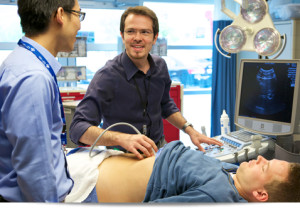
The ultrasound technician after that moves the particular transducer around till a desirable image can be seen and interpreted within the monitor. Technicians are usually trained to look for any kind of abnormalities that typically the ultrasound may pick up and note these for that patient’s physician. The required images are after that saved for the physician’s viewing later. Following the procedure, the skin gels is easily wiped from the patient’s skin.

Even though a noninvasive treatment that requires no distress aside from the frigidness from the ultrasound gel, ultrasounds need to be used with extreme caution in certain situations, like in obstetrics. It is necessary that pregnant women make use of the technology available today to guarantee the health of their child, but studies have demonstrated that extreme ultrasound procedures, such as within the vanity ultrasound video clips being offered nowadays, can lead to developmental problems, based on the U. S. As well as Drug Administration. However, as long as the process is performed by a certified ultrasound technician and below doctor’s orders, the advantages far outweigh the potential risks.

Ultrasound – Finally the Good Hits the Bad

Healthcare imaging has gone through a sea improvement through the years, and now all of us have a variety of techniques that consider our entire body and help physicians diagnose what is incorrect with us. Specific types of injuries and illnesses require certain types of radiology techniques, yet ultrasound examinations appear to score over the relax in situations exactly where it can be used. Which is because:

Ultrasound Benefits:

  • Ultrasound is really a noninvasive procedure and you also don’t have to be able to deal with this and discomfort associated with tubes together with scopes being placed into the body.
  • It’s not since expensive as other styles of medical image resolution because the machines is cost effective.
  • It’s convenient and quick.
  • It’s transportable since the device is light enough to become moved from place to place.
  • It is usually accessible at your own doctor’s office, hence saving you the trouble associated with going to a several place for your imaging requirements.
  • It is safe because it does definitely not use dangerous radiation such as X-rays or even MRI tests and therefore, is the very best kind of examination for expecting a baby mothers.
  • It can get used to deliver therapeutic massages and physical therapy.
  • It really is even used to split renal stones in a pain-free, noninvasive procedure.
  • It will not use any chemical substances to visualize your own innards.
  • It uses current imaging (images show up as soon as the particular transducer is positioned on skin) which allows the radiologist to find the movement of tissue.
  • The real-time image resolution also enables ultrasound to be utilized to guide biopsies.
  • Doppler ultrasounds may be used to evaluate blood flow within blood vessels and even in strong organs and world.
  • Intravenous ultrasound can be used to evaluate often the internal parts of blood vessels.
  • Concentrated ultrasound is utilized to execute surgeries for remedying of tumors such as fibroids in the particular uterus.

For the negative side though:

  • Ultrasound can be utilized only to picture internal organs which usually are made from soft tissues.
  • This cannot be utilized to picture hard structures such as the bone and internal organs filled with air such as the lungs and feces.
  • The images are usually little difficult to translate unless you’re a tuned radiologist.
  • The low quality makes it difficult to obtain detailed anatomical details.
  • It’s hard to obtain decipherable pictures within obese people since fat is a bad transmitter of audio.
  • The variety of eyesight is limited.
  • The presence of atmosphere or gas the actual quality of pictures poorer than they are definitely.
  • When used for a long time at high intensities, that can cause the particular tissues to turn into warmed.

The positive factors of ultrasound outweigh the downsides, and this is why ultrasound continues to be a popular method to produce professional medical images of humans.